All posts by admin

Diastema

6922

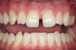
Whether you love it or hate it, that space between your teeth has a name. A diastema is a gap between two teeth. Many celebrities are famous for their midline diastema, or space between their two upper front teeth.Diastemas are extremely common, especially among children. A diastema is a natural part of a child’s development and may correct on its own. In fact, up to 97 percent of children have diastemas, and that number significantly decreases as children grow and these spaces close up naturally. If a diastema remains after the eruption of adult teeth, it will become permanent and can only be corrected with professional diastema treatment.


Diastema Causes

There are several reasons that permanent diastemas form. A diastema is often the result of a discrepancy between the size of the jaws and the size of the teeth. Crooked teeth usually come from overcrowding, where the teeth are too big for the jaw. The opposite is true for a diastema — teeth that are too small for the jaw may have gaps between them. Diastemas may also be caused by missing teeth, undersized teeth or bad oral habits, such as excessive thumb-sucking.A midline diastema can also be caused by a large labial frenum. Frenum is the tissue that connects your lips and gum.

Diastema Closure :- Diastemas usually cause no complications to your dental health, but many people choose diastema closure for cosmetic purposes. There are several types of diastema treatment available today.

Dental Braces — Most diastemas require a full set of dental braces and retainer therapy, as moving one tooth can affect the placement of the rest.



Torus Palatinus

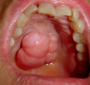
Torus palatinus is a benign peculiarity wherein there is a bony outgrowth on the palate. It is a normal bump on the roof of the mouth. It is one of the most common exostosis involving the oral cavity. It is not cancerous, nor is it painful or sore. It comes in different shapes: nodular, spindle-like, lobular, or irregular. It is hard upon palpation and radiography shows opacity on this area due to the bone’s density. Since it is normal, there is no symptom associated with it.

      Torus Palatinus Causes

torus-palatinus-picturesGenetic and ethnic factors: Development of torus palatinus in parent/s poses a 40-64% chance that their offspring will have it too. If neither of the parents has it, there is a       5-8% chance that a child will have it.                                   Torus palatinus occurs in 20% of the general population. Torus mandibularis occurs in 6-40%.It mostly appears after puberty. It is generally seen among adults. For children in 5-15 age groups, there is 30.9% occurrence and it was shown that females are more likely to develop it. Torus palatinus is more common among females. Torus mandibularis is more common among males. Torus palatinus does not need to be treated since its essential features appear to be normal. It is simply an anatomical variance that does not affect everyone of us. Surgical removal offers a solution if torus palatinus causes a problem in the placement of dentures or dental prosthesis.

Sialadenitis

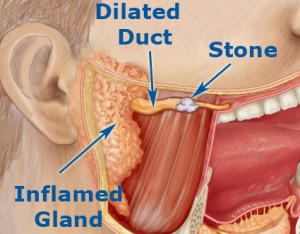
Sialadenitis is the infection of the salivary glands caused by a bacteria or virus. The parotid, located in front of the ear and submandibular, placed below the chin are the glands, which are affected most commonly. They cause ache, tenderness, redness, and steady concentrated inflammation of the region affected. It can be either acute or chronic, and is frequent among adults with salivary gland stones. However, it can occur to anyone, including infants. If not treated well, Sialadenitis can progress into a severe infection.   Causes of Sialadenitis. Bacteria cause most of the acute cases, especisalivary-gland-stones1ally staphylococcus infections. Ignorance of oral hygiene is a major reason for this infection. Apart from these, infections like HIV, herpes, and mumps can also lead to the swelling of the salivary gland. Chronic Sialadenitis is often caused by salivary stones, which are nothing but deposits of calcium and different minerals that get accumulated in the glands and lead to blockages. Although less frequent, a person can also develop this infection due to the result of the immune system responding to other drugs given to treat different glandular cases.

Dry socket(Alveolar osteitis)

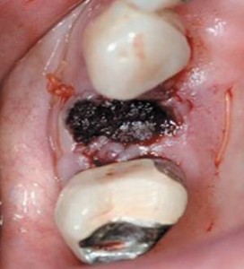
The socket is the hole in the bone where the tooth has been removed. After a tooth is pulled, a blood clot forms in the socket to protect the bone and nerves underneath. Sometimes that clot can become dislodged or dissolve a couple of days after the extraction. That leaves the bone and nerve exposed to air, food, fluid and anything else that enters the mouth.

Symptoms may include:dry

Dull, aching, throbbing pain in the area of the socket, which is moderate to severe and may radiate to other parts of the head such as the ear, eye, temple and neck. The pain normally starts on the second to fourth day after the extraction and may last 10–40 days. The pain may be so strong that even strong analgesics do not relieve it,Intraoral halitosis,Bad taste in the mouth.

Dental Abscess with Facial Cellulitis

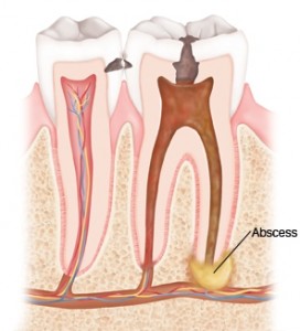
A dental abscess is an infection at the base of a tooth. It means a pocket of pus has formed at the tip of a tooth root in your jaw bone. If the infection isn’t treated, it can spread to the gum near the tooth. This causes swelling and pain. More serious infections spread to the face. This causes your face to swell (cellulitis). This is a very serious condition. Once the swelling begins, it can spread quickly .A dental abscess usually starts with a crack or cavity in a tooth. The pain is often made worse by drinking hot or cold beverages, or biting on hard foods. The pain may spread from the tooth to your ear or the area of your jaw on the same side.

115953_1.psd

Aerodontalgia

Pain in the teeth that results from a change in air pressure (as in flying or mountain climbing) .  Aerodontalgia affects some person who experience pain in the tooth during high altitude flight or during deep sea diving. At ground level the tooth is coshutterstock_12851194mpletely asymptomatic. In some cases the pain may not start readily during flight or during diving, instead it may occur few hours or days later .The condition may be related to  subclinical pulpitis. Sometimes similar problem may happen in an endodontically treated tooth with improper obturation of the canal. The entrapped air in an improperly obturated canal may expand during flight or driving due to alteration in atmospheric pressure. This applies pressure in periapical nerve bundle and produce pain.

TONGUE THRUST

Tongue thrust, also known as reverse swallow or deviate swallow, is a condition in which the tongue is pressed up against the teeth or between them while gulping. In many cases, this condition leads to the movement of the tongue incorrectly to the sides, instead of forward to the teeth. This improper positioning of the tongue can occur even as the tongue is at rest, and in this case, it will lie too far forward.

Types of Tongue Thrust

Tongue thrust is an orofacial myofunctional disorder (OMtongue thrust imageD), having many different types.

  • Improper closing of the lips, also known as anterior open bite.
  • Anterior thrust refers to the case when the upper incisors are stuck out.
  • Bilateral anterior open bite is the situation in which the children have a tongue that is larger than normal and when the bite is closed, the molars are the only teeth, which are in contact.


Bulimia

The dental changes seen in many people with bulimia are often recognizable. Frequent vomiting may cause your salivary glands to swell and the tissues of your mouth and tongue to become dry, red and sore. People with bulimia may have chronic sore throat and small hemorrhages under the skin of the palate.
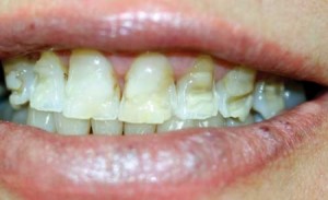
Frequent vomiting can erode your tooth enamel, especially on the tongue side of the upper front teeth. This sharply increabulimia imageses the risk for decay in these areas and can make these teeth sensitive to temperature. Severe erosion can lead to changes in your bite, or the way your upper and lower teeth come together. Your back teeth can be reduced in size and some teeth can even be lost eventually. Tooth erosion can take about three years to become obvious, but not all bulimics experience it.
Many people with bulimia may be malnourished, which can cause anemia, poor healing and increase the risk of periodontal disease.

Tetracycline stain

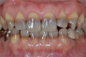
Tetracycline-stains pickTetracycline teeth stains develop on permanent teeth while they are still forming under the gum line. During development, the drug becomes calcified in the tooth, generating tetracycline tooth stains. Children are susceptible to tetracycline tooth stains from the time they are in utero until the age of 8. Since teeth start to develop before we’re even born, pregnant women should not take tetracycline to prevent the possibility of the drug affecting the unborn baby’s oral health.Gray or brown in color, these deep, dark stains either cover the entire tooth or appear as a pattern of horizontal stripes. Because they form during tooth development, tetracycline teeth stains are embedded in the tooth’s enamel and inner layers. This classifies them as intrinsic stains — discoloration that comes from inside the tooth. Although harmless, tetracycline stains are obvious and permanent, often causing embarrassment and low self-esteem in those who suffer from them.

BOOK AN APPOINTMENT