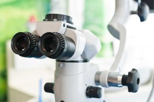
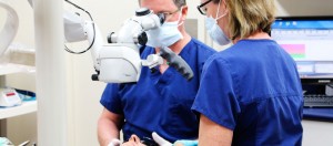
From guessing dentistry to precision dentistry has changed the way dental treatments were done. Application of microscope in the field of dentistry has been very recent. It lets the dentist to visualize familiar structure in the totally different way, with finest details that were earlier concealed from the naked eyes. It is an indispensable device which is connected with high resolution video camera to project the magnified images on the LCD monitor that clients can also see. It allows dentist to get a better view of the patient’s oral cavity, thereby improving the overall results of the treatment, while limiting trauma and increasing precision.
A microscope in dentistry has wide spectrum use, idea of this technology is to remove as little of the natural teeth as possible while removing all of decay, it permits complex system of canals to be recognized, managed and identify in a safe manner. It also makes easier to handle cases that in past would be impossible to resolve without magnification, such as retrieval of broken pieces of an instruments during dental procedures from root canal, management of stones and calcification in the root canals, visualization of accessory or multiple canals, missed canals in a tooth and a better prognosis for re-treatment’s.
It also facilitates the on demand documentation of the treatment (both video and photographic)
How does microscope enables dentist to provide better patient care?
The microscope gives increased precision and a higher level of confidence that all decay has been removed. Importantly, it enables better diagnosis and effectively communicates these to patients.
What are its other advantages?
The biggest advantage is during re-treatment. To perform a re-treatment can be as simple as the removal of gutta -percha from a poorly obturated canal to more complex, delicate and time consuming procedures, like removing screw posts, separated instruments, silver points, amalgam pins, carbon fiber posts, or re- pairing a perforation or obturating an im-mature open apex.


