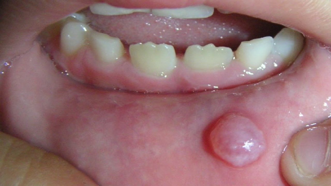
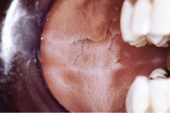
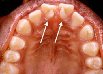
Dental plaque


dental-plaque

Dental plaque, also known as tooth plaque, microbial plaque and dental biofilm, is a soft, sticky film that builds up on your teeth. Plaque is an extremely sticky, colorless to pale yellow deposit of biofilm that regularly forms on your teeth. When saliva, food, and fluids combine they produce bacteria deposits, which collect where the teeth and gums meet. Plaque contains bacteria, which produce acids that attack your tooth enamel and can damage your gums. If not treated, the damage could become permanent.  It contains millions of bacteria that feed on the food and drinks you eat

What Causes Plaque and Why Is It Harmful?

Plaque develops when foods containing carbohydrates (sugars and starches), such as milk, soft drinks, raisins, cakes, or candy are frequently left on the teeth. Bacteria that live in the mouth thrive on these foods, producing acids as a result. Over a period of time, these acids destroy tooth enamel, resulting in tooth decay. Plaque can also develop on the tooth roots under the gum and cause breakdown of the bone supporting the tooth.

So why is plaque a problem?

If you don’t take the steps needed to prevent and get rid of plaque, it may lead to:-

Cavities: the acids produced by the bacteria in plaque can cause low pH level and can eat away at your tooth enamel.

Gingivitis: Accumulation of plaque bacteria can cause inflammation of the gums.

Bad Breath: Plaque buildup from poor dental hygiene can also cause your breath to smell bad.

How to prevent tooth plaque:-

  •  Visit your dentist every 6 months. Adults who see their dentists regularly are less likely to have plaque related dental diseases.
  • Floss daily to remove food particles and plaque between teeth. To prevent the buildup of plaque, be sure to floss once a day  and brush your teeth at least twice a day with toothpaste.
  • If you have electric toothbrush, and remember to replace the head every 3 months. Use a quality toothbrush, or as soon as the bristles look worn.
  • Avoid smoking to reduce the risk of increased plaque and tartar.
  • Use a mouthwash such as Multi-Protection Mouthwash to help prevent plaque buildup on teeth to your routine against plaque and gingivitis.

images


Dental Cyst

cycst

What Is a Dental Cyst?

A dental cyst is very much like a closed sack. What is inside the sack can differ from patient to patient. Some cysts are comprised of air, some of soft tissue, and others are filled with fluid that can build up next to and around teeth. A dental cyst left to grow without treatment can lead to severe problems for your oral health.

Where Are Cysts Found?

There are various types of dental cysts.

Periapical cyst:-These cysts form at the root tip, and are typically caused by trauma, a crack in the tooth, or decay that has infected or killed the nerve (pulp) of the tooth.

Dentigerous Cysts: These cysts grow around unerupted or partially-erupted teeth, particularly wisdom teeth.

Keratocysts: These cysts typically form due to trauma or genetics. These cysts are aggressive and exhibit a high rate of recurrence, even after surgical removal.
Periodontal Cysts: These types of cysts are caused by advanced periodontal or gum disease, and thus are bacterial in nature.

Potential Oral Health Problems

A cyst left to grow can create unwanted pressure on teeth that can have the opposite effect on your smile. For example, a worsening cyst can eventually affect your bite by moving teeth, change the way you chew and digest food, and weaken the jawbone. In extreme cases of undetected cysts, the jawbone can actually be put at risk for fracture, among other complications.

Furthermore, a dental cyst can become infected and can eventually develop into an abscess, or pus-filled sack, which can spread the bacterial infection throughout your body.

Now that we have determined that cysts are bad, how will your dentist treat the cyst found in your mouth?

How to Treat Dental Cysts

The dental cyst will first require a thorough examination by your dentist or maxillofacial surgeon. The type of treatment used will depend on the type of cyst as well as where it’s located.

A cyst that forms on the root of a tooth might require endodontic or root canal treatment in order for the cyst to begin the healing process.

Leukoedema

846

Leukoedema is the normal anatomic variant of the oral mucosa which has clinical appearance similar to potentially malignant white lesions such as leukoplakia and lichen planus. Other lesions which closely mimic leukoedema are white sponge nevus and cheek bite.

Leukoedema is a harmless white lesion of the oral cavity which mimics premalignant lesion more so when it is associated with smoking.


Signs and symptoms

There is a diffuse, gray-white, milky opalescent appearance of the mucosa which usually occurs bilaterally on the buccal mucosa. The surface of the area is folded, creating a wrinkled, white streaked lesion. Apart from the appearance, the lesion is entirely asymptomatic.

Causes

The cause is unknown, but it is thought to be caused by intracellular edema of the superficial epithelial cells coupled with retention of superficial parakeratin. Although leukoedema is thought to be a developmental condition, it may be more common and more pronounced in smokers, and becomes less noticeable when smoking is stopped. Smoking cannabis is known to be linked to this condition. It may also develop in areas subjected to repeat subclinical irritation, caused by low grade irritants such as spices, oral debris or tobacco.

Diagnosis

Clinically

Treatment

No treatment is required for leukoedema.


Mouth sores(Ulcers)

mouth ulcer 2

Mouth ulcers are painful areas in the mouth and gums.

While mostly harmless, mouth ulcers can be extremely uncomfortable and make it difficult for some people to eat, drink, and brush their teeth.

Mouth ulcers range in size, and the exact symptoms of the mouth ulcer will depend on what type of ulcer a person has.

Fast facts on mouth ulcers:

  • Most mouth ulcers are recurring
  •  Acidic foods can aggravate mouth ulcers.
  • It is important to note that a doctor or dentist should examine any new ulcer and any ulcer lasting longer than 3 weeks.
  • For most people, mouth ulcers will clear up within 2 weeks.

Causes

The exact cause of mouth ulcers is still not known and varies from person-to-person. Still, there are some common causes and several factors that may aggravate mouth ulcers, including the following:

  • Biting the tongue or inside of the cheek
  • Braces, poor-fitting dentures, and other apparatus that may rub against the mouth and gums
  • A deficient filling
  • Stress or anxiety
  • Hormonal changes during pregnancy, puberty, and menopause
  • Medications including bit-blockers and pain killers
  • genetic factors


Symptoms of minor and major ulcers include:

  • one or more painful sores that may appear on the cheeks, the roof of the mouth, or the tongue
  • the appearance of round lesions that have red edges and are yellow, white, or gray in the middle

During more extreme outbreaks of mouth ulcers, some people may experience fever, sluggishness, and swollen glands.

Treatment

In many cases, the pain and discomfort from mouth ulcers will lessen in a few days and then disappear in about 2 weeks with no need for treatment.

For people with much more painful or frequent recurrence of mouth ulcers, a dentist may prescribe a solution to reduce swelling and lessen pain.

Also, a dentist may prescribe an antimicrobial mouthwash or an ointment to be applied directly to the infected patch. This can help to ease discomfort. Various mouth ulcer treatments are also available to purchase

  • Avoid spicy and sour foods until the ulcers heal.
  • Drink plenty of fluids.
  • Regularly rinse your mouth out with warm, slightly salted water.
  • Keep your mouth clean.
  • Use an alcohol-free medicated mouthwash
  • Use a topical steroid mouthwash or ointment – generally prescribed by your dentist or oral medicine specialist.


Tongue Thrust

tongue

Tongue thrust, also known as reverse swallow or deviate swallow, is a condition in which the tongue is pressed up against the teeth or between them while gulping. In many cases, this condition leads to the movement of the tongue incorrectly to the sides, instead of moving forward to the teeth. This improper positioning of the tongue can occur even as the tongue is at rest, and in this case, it will lie too far forward.

Types of Tongue Thrust

Tongue thrust is an orofacial myofunctional disorder (OMD), having many different types.

  • Unilateral thrust when the tongue pushes unilaterally to the sides between the back teeth during swallowing
  • Anterior thrust refers to the case when the upper incisors are extremely protruded and lower lip is pulled in by the lower lip.
  • Bilateral thrust, when the tongue pushes between the back teeth on both sides during swallowing. Large tongue can also be noted

Tongue thrust in babies

In babies who are breastfed or bottle-fed, tongue thrust is normal. As the child gets older, their swallowing and speaking pattern should evolve.

However, some types of bottle nipples — and prolonged use of a bottle — can lead to an abnormal tongue thrust that lasts past the infant stage and into early childhood.

There are several other potential causes for tongue thrust that begin in infancy. Some of these include:

  • Long-term sucking habits that influence the tongue’s movement, like sucking of the thumb, fingers, or tongue
  • Allergies accompanied by chronically swollen tonsils or adenoids
  • Tongue-tie, where the band of tissue beneath the tongue is tight or short
  • A swallowing pattern known as reverse swallow

Tongue thrust has a number of telltale signs that manifest in children who have developed the pattern. These can include:

  • Tongue is visible between the teeth. The tip of the tongue sticks out between the teeth, whether the child is resting, swallowing, or speaking.
  • Mouth breathing.
  • Inability to close the lips completely. This could be due to a structural abnormality or habit.
  • Open bite. This occurs when the front teeth don’t meet when the teeth are closed.
  • Slow, fast, or messy eating.
  • Speech impediment. Lisping of s and z sounds is common.

Treatment of Tongue thurst

Tongue thrust often corrects with time or at the age of 8-9 years. Self-correction is due to musculature Balance during swelling.

  • Orthodontic treatment can be carried out.
  • Speech therapy is indicated.
  • If an associated habit is present like thumb sucking than it must be treated first.
  • Training the tongue for correct swallow & posture.
  • Use of appliances to correct position of tongue.
  • Correction of Malocclusion.

Leukoplakia


Leukoplakia is a reaction to a chronic irritation, like rough teeth, badly fitting dentures, smoking, and smokeless tobacco. It can show up as white patches or plaques in the mouth, is usually painless, and can’t be scraped off. Leukoplakia can also be a precancerous condition. Persistent patches or other changes in your mouth. Mild leukoplakia is usually harmless and generally goes away on its own. More serious cases may be linked to oral cancer.

Symptoms of Leukoplakia:-

This condition is marked by unusual looking patches inside the mouth. These patches can vary in appearance. Generally, patches resulting from leukoplakia have the following features:

  • white or grey color
  • thick, hard, and raised surface
  • hairy (hairy leukoplakia only)

Rarely, the patches have red spots. Redness may be a sign of cancer.                           Leukoplakia most often occurs on the tongue. However, cheeks and gums are also vulnerable. The patches may take several weeks to develop, but they are rarely painful.      Causes:­-Smoking is the most common cause. Chewing tobacco can also cause leukoplakia. Biting the cheek, rough uneven teeth  and dentures (especially if improperly fitted).   


What Causes Leukoplakia?

  • Irritation from rough teeth, fillings, or crowns, or ill-fitting dentures that rub against your cheek or gum
  • Chronic smoking, pipe smoking, or other tobacco use
  • Sun exposure to the lips
  • Oral cancer (although rare)
  • HIV or AIDS

What Are the Symptoms of Leukoplakia?

The presence of white or gray colored patches on your tongue, gums, roof of your mouth, or the inside of the cheeks of your mouth may be a sign of leukoplakia. The patch may have developed slowly over weeks to months and be thick, slightly raised, and may eventually take on a hardened and rough texture. It usually is painless, but may be sensitive to touch, heat, spicy foods, or other irritation.

How Is Leukoplakia Diagnosed?

Your dentist may suspect leukoplakia upon examination; however, a biopsy will likely be taken to rule out other causes, such as oral cancer. During the biopsy, a small piece of tissue from the lesion will be removed to be examined in a lab. A numbing agent will be used so that you will not feel any pain.

Treatment for Leukoplakia?

Treatment for leukoplakia, if needed, involves removing the source of irritation.

For example, if leukoplakia is caused by a rough tooth or an irregular surface on a denture or a filling, the tooth will be smoothed and dental appliances repaired.

If leukoplakia is caused by smoking, you will be asked to minimize or stop smoking or using other tobacco products.

leuko


DIFFERENCE BETWEEN ABFRACTION, ABRASION, EROSION AND ATTRITION

DIFFERENCE BETWEEN ABFRACTION, ABRASION, EROSION AND ATTRITION

Abfraction, abrasion, erosion and attrition  all involve some tooth damage, but at different locations on the tooth. While they have varying causes, they can interact and create a bigger problem. It’s possible to have abfraction, abrasion, erosion and attrition at the same time.

Abfraction

Abfraction is a wedge-shaped flaw on the tooth at the point it meets the gumline.

It’s caused by friction and pressure on the tooth and gums, which causes the neck of the tooth to start breaking off.

Abrasion

Abrasion is likely to be found on the teeth closest to your cheeks, also known as the buccal side. Unlike the V-shaped appearance of abfraction, the damage caused by abrasion is flat.

Abrasion is caused by friction from foreign objects, such as pencils, fingernails, or mouth piercings. Using a hard toothbrush, abrasive tooth products, and improper brushing technique can also lead to abrasion.

Erosion

Erosion is the general wearing away of tooth enamel. Teeth may have a more rounded appearance, with hint of transparency or discoloration. As erosion progresses, you can start to see dents and chips in the teeth.

Unlike abfraction and abrasion, erosion is more of a chemical process, happening on the surface and subsurface of the teeth. It’s caused by high acid levels in the saliva. This may be due to acidic foods or drinks, dry mouth, or health conditions that cause frequent vomiting.

Attrition

Dental attrition is a type of tooth wear caused by tooth-to-tooth contact, resulting in loss of tooth tissue, usually starting at the incisal or occlusal surfaces. Tooth wear is a physiological process and is commonly seen as a normal part of aging.

blooog pics

Tooth Abfractions

afraction

Abfraction lesions on teeth are small notches caused by stress (forces) on your teeth. Biting, chewing, clenching and grinding put pressure on your teeth. Over time, this pressure can cause cracks and splits in the outer layer of your teeth near the gum line. Such lesions are fairly common in adults. Older adults are especially likely to have them. They occur more often in the back teeth, called premolars and molars. But they can occur in the front teeth as well and appear as V-shaped or wedge-shaped notches. Abfraction lesions don’t usually hurt but if the deeper layers of the tooth are involved, sensitivity might occur. These lesions do not heal over time. Some may worsen, and if left untreated, can cause the tooth to fracture. In order to prevent this, your dentist might recommend fillings of these lesions. This will not only strengthen the tooth, but also decrease the sensitivity if present.

What are the symptoms of abfraction?

You might first become aware of abfraction when you get food stuck in the wedge or when you flash a big smile. You might even be able to feel it with your tongue.

Abfraction is usually painless, but tooth sensitivity can become a problem, especially where heat and cold are concerned.

You may never develop other signs or symptoms, but if the damage continues, it could lead to:

  • worn and shiny facets on the tooth, known as translucency
  • chipping of the tooth surface
  • loss of enamel or exposed dentin

What causes abfraction?

Abfraction is caused by long-term stress on the teeth. This can happen in a variety of ways, such as:

  • bruxing, also known as teeth grinding
  • misalignment of the teeth, also called malocculusion
  • mineral loss due to acidic or abrasive factors

How is abfraction treated?

The diagnosis can usually be made on clinical examination. Tell your dentist about any health conditions or habits that can affect the teeth. Some examples of this are:

  • habitually clenching or grinding your teeth
  • eating disorders
  • a highly acidic diet
  • wrong brushing techniques
  • medications that cause dry mouth

Your doctor will recommend treatment based on the severity of your symptoms and whether you have co-existing dental problems. You might also want to consider how it affects your smile and ability to keep your teeth clean.

The damage can’t be reversed, but you can ease tooth sensitivity, improve appearance, and help prevent future damage. Some treatment options are:

  • Fillings.
  • Nightguard.
  • Toothpaste.
  • Orthodontics.


Midline Diastema


midline

Whether you love it or hate it, that space between your teeth has a name. A diastema is a gap between two teeth. Many celebrities are famous for their midline diastema, or space between their two upper front teeth.Diastemas are extremely common, especially among children. A diastema is a natural part of a child’s development and may correct on its own. In fact, up to 97 percent of children have diastemas, and that number significantly decreases as children grow and these spaces close up naturally. If a diastema remains after the eruption of adult teeth, it will become permanent and can only be corrected with professional dental treatment.

Diastema Causes

There are several reasons that permanent diastemas form. A diastema is often the result of a discrepancy between the size of the jaws and the size of the teeth. Crooked teeth usually come from overcrowding, where the teeth are too big for the jaw. The opposite is true for a diastema — teeth that are too small for the jaw may have gaps between them. Diastemas may also be caused by missing teeth, undersized teeth or bad oral habits, such as excessive thumb-sucking.A midline diastema can also be caused by a large labial frenum. Frenum is the tissue that connects your lips and gum.

Diastema Closure :- Diastemas usually cause no complications to your dental health, but many people choose diastema closure for cosmetic purposes. There are several types of diastema treatment available today.

Dental Braces — Most diastemas require a full set of dental braces and retainer therapy, as moving one tooth can affect the placement of the rest.

Cosmetic restoration—Esthetic composite resins generally used to close midline diastema especially in adult patients. It requires a gradual composite build-up on the mesial side of the tooth and stripping of distal surface of the tooth in order to achieve a natural shape and size of the tooth.


Dental Implant vs Bridge vs Denture


A Dental implant is a titanium post (like a tooth root) that is surgically positioned into the jawbone beneath the gum line that allows your dentist to mount replacement teeth.

implany

A Dental bridge is a false tooth that is held in place by the adjacent teeth on either side of the gap. Although artificial tooth  can be made from a variety of materials such as gold, typically they’re made from porcelain to aesthetically blend in with your natural teeth.

shutterstock_713159425

Dentures are prosthetic devices constructed to replace missing teeth, and are supported by the surrounding soft and hard tissues of the oral cavity. Dentures are replacements for missing teeth that can be taken out and put back into your mouth.There are two main types of dentures: full and partial.

E-Dent-and-E-Denture-e1498856989959


Basis Dental Implants Dentures Dental Bridges
Performance Look and like natural teeth Function similar to  natural teeth Look and like natural teeth
Comfort Secure confident smile Poorly fitting denture can irritate gum tissue Sensitivity in supporting teeth
Durability Create normal bite force Reduce bite force Vulnerable to bite force
Bone loss Maintain jaw bone Causes loss of jaw bone Doesn’t prevent bone loss
Impact on natural teeth Doesn’t impact on natural teeth Partial denture can wear and increase cavities Require modification of natural teeth
Longevity Provide long term stability Less than a year Doesn’t stable more than 8 years Can last 10-20 years
Appearance Eat your favourite food Limited food choice Eat only limited food
Cost More expensive Less expensive More expensive
Time Take one month or one year depend on the mouth condition Less than one month Less than one month
Hygiene Brush, floss and see dentist like natural teeth Must be removed to clean and should not be worn while sleeping Cleaning under the bridges

BOOK AN APPOINTMENT